

Left Brain, Right Brain
Alumnae Taps Into the Educational Power of Science + Art
By Gaye Hill
Left Brain, Right Brain
Alumnae Taps Into the Educational Power of Science + Art
By Gaye Hill
Even as a toddler, Joanna Cox, ’17, was intensely creative. So it’s no surprise that before she arrived at Meredith, she had already completed four years of study in fine arts, having attended a performing and visual arts high school.
 Once at Meredith, when she was deciding on her major, she felt ready for a change. So rather than continuing her study of art, she decided to major in biology, which had always been another area of interest.
Her intention was to earn a Ph.D. and pursue a career in research. To that end, she held an internship at the National Institute of Environmental Health Sciences in the Research Triangle for two years.
“I loved research, and I loved working in a lab,” said Cox. “But just looking at how someone typically progresses within the research industry – the more senior you become, the less hands-on work that you’re doing, and much of the job involves grant writing. I wasn’t really a fan of that aspect of the job.”
Cox started exploring other career options that might allow her to combine her two passions: science and art. She came across medical illustration and discovered there were graduate programs that specialize in this unique field.
Mentors and Internships
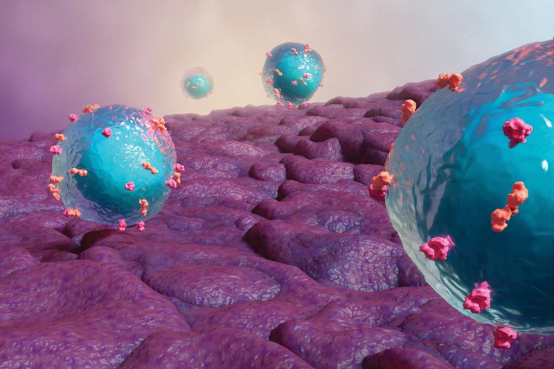
Illustration of CAR T cells invading a tumor environment.
Around that time one of her mentors at Meredith, Associate Professor of Biological Sciences Maria Pickering, connected her with a colleague at the North Carolina Museum of Natural Sciences who was doing fieldwork studying crayfish.
“I had the opportunity to do a scientific illustration internship in that lab,” she said. “So I was doing some light microscopy and sketching microbes that she found on creeper specimens. I was also doing plate photography, taking photographs of the different features of crayfish that she would use to define characteristics within species.”
Another mentor, Professor of Biological Sciences Jason Andrus, supported Cox as she developed an interactive lab manual that used QR codes to provide short videos demonstrating lab techniques.
“She was very motivated, talented, and curious,” said Andrus in describing Cox. “She was not afraid of challenges and really took ownership of her educational experience.”
Andrus said Cox worked hard to pursue a medical illustration career path, which is a niche field and one that is extremely competitive.
“It requires excellent artistic ability as well as the ability to understand biological principles,” said Andrus.
Well-Prepared for Graduate School
After graduating from Meredith, Cox studied medical illustration at the Rochester Institute of Technology (RIT). She thrived in her program, in part thanks to the support of her professors encouraging her to go for what she wanted.
“I’ve always been a very studious, organized person so the transition to graduate school wasn’t difficult for me. I had an independent drive to achieve the next step in my career.”
With a cohort of classmates who were all from out of state, she formed strong bonds with those in her program and felt supported by her professors, who she described as “great humans and fantastic teachers.”
She also immediately took to her studies.
“The content of the classes was so interesting,” said Cox. “In human gross anatomy, we were dissecting cadavers and really getting to know and understand where things lie in the body so we could more accurately illustrate. That was totally awesome. Outside of medical school, a lot of people don’t really get to experience that.”
They would also live-sketch while observing surgeries and then come back and create a more substantive piece using their sketches. Cox enjoyed the opportunity to produce both 2D and 3D illustrations, which drew on her fine arts background while adding new technical modeling and animation skills.
Communicating Science Through Art
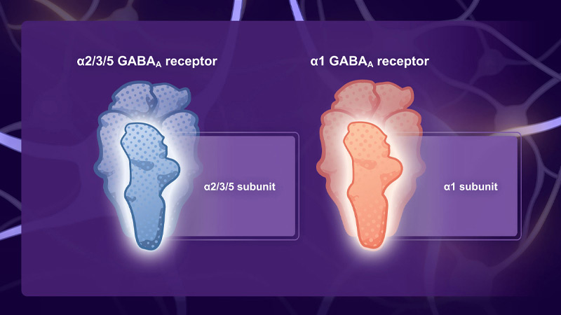
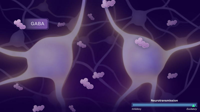
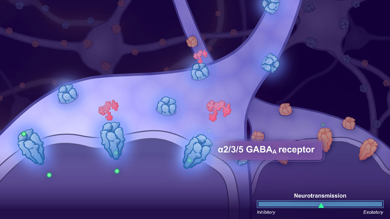
Three excerpts from the GABA modulation video courtesy of Engrail Therapeutics.
“The ultimate goal of our industry is to beautifully and accurately describe scientific content,” said Cox. “Obviously the work you’re producing is largely dependent upon the target audience. If your target audience is healthcare professionals, then you’re going to include more scientific detail. For a patient audience, you would want to minimize the amount of overwhelmingly specific details you’re sharing with them.”
Cox noted that she works in medical communications but the industry is diverse, with illustrators found in research institutions such as UNC-Chapel Hill and the Howard Hughes Medical Institute as well as publishers such as Science magazine. She has colleagues who work for hospitals where they design illustrations to create pamphlets for patients to help them understand surgery they’re undergoing or a diagnosis.
“It can range between truly educational and really beautiful,” she said.
Cox met Peter Domiano in graduate school. They now collaborate on scientific visuals at Boldscience, where Domiano is the lead medical illustrator.
“Joanna has worked on many Mechanism of Action illustrations and videos for large pharmaceutical and biotechnology clients,” said Domiano. “This content has helped teach healthcare providers, patients, and internal staff about emerging treatments for diseases.”
He noted that she is an expert at using color and design to guide the viewer through scientific content.
The ultimate goal of our industry is to beautifully and accurately describe scientific content.”
“These color and design skills give a crisp and clean look to her illustrations, which stands out in our field,” he said. “Combined with her extensive scientific background, her work is clear and effective in teaching the intended audience.”
An active national association and conferences foster strong collaboration among colleagues, who are eager to learn about advances in technology and apply them to their work. Such connections also lead to a certain brand of humor.
“There are running jokes in our field,” she said. “Sometimes people run into illustrations that have a backward brain so the cerebellum is in the front and it’s supposed to be in the back. Or DNA is twisted the wrong way. You send it to your friends in the medical illustration field and they’re like, ‘oh, that is hilarious.’”
Jokes aside, there are extensive processes in place to ensure medical illustrations are correct. Cox’s work setting comprises a scientific team and a creative team. Both teams review all illustrations carefully before they are provided to a client.
“Obviously, as a medical illustrator, you need to make sure your work is accurate. If it’s not, that completely takes away the integrity of the piece,” she said.
Bridging the Scientific Knowledge Gap
According to Cox, one of her primary goals in going into this field was to help bridge the gap between scientists and the public.
“COVID-19 was an unbelievable example of science disinformation or lack of understanding,” she said. “But if you talk to a research scientist about their research and you’re a layperson, chances are you’ll understand about three words they’re telling you because they are so excited about their work they don’t realize they’re talking in such a technical way and using jargon.”
Cox said the Centers for Disease Control (CDC) has medical illustrators on their staff and noted the ubiquitous virus particle with the spikes was created by one of their resident medical illustrators.
She is excited about emerging developments in the field, too. At a recent conference, she heard from a speaker who uses MRI and CT scan data to create 3D models of tumors. Such models are used to educate the patient on where their tumor is located as well as by surgeons who are operating on the patient to help determine the least invasive approach.
That kind of exciting technology is one reason Cox loves her job.
“I am immensely grateful that I found this field because it quite literally marries my two biggest passions.”